Saturday, 30 August 2008
Bioreactor-in-progress...
Photo Credits: matt Johnson
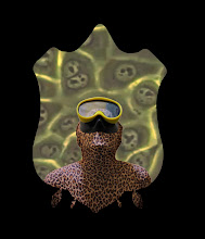
Captain Science Matt Johnson (mattmjohnson.com) and I imitating bad chemistry experiments.
Saturday, 16 August 2008
bioreactor in mini ecosystem
This is an initial concept sketch of the mini ecosystem which will contain the bioreactor, in which the tissue taken from my thigh will be grown into sculptural form.
The top vessel is the bioreactor. the second tank will be a aquarium, and the bottom one a plant tank. All constructed from glass.
Image+ Design: Matt Johnson
Image + Design: Matt Johnson
My current Residency at SymbioticA (www.symbiotica.uwa.edu.au) involves designing the bioreactor, in collaboration with Matt Johnson an industrial design student from the RCA in London, and Oron Catts as part of The SymbioticA Research Group.
While here at symbi i'm also working on a project involving thetissue cuturing of excess surgical tissue from cosmetic surgery patients, and doing HIV/Lentivirus research following from Go Forth an Multiply, mentioned below.
Slip me some skin is a project involving the culturing of my own tissue outside my complete living body. This project involved indergoing a tissue biopsy, to remove primary tissue from my body, to be cultured within The University of Tasmania's School of Medicine's Pathology Dept. On the 18 March at 4pm I met my appointment at an undisclosed surgery, where an eliptical piece of tissue 3cm x 1cm was removed from my inner thigh.
Video footage of this process was taken and is being compiled into a video piece.
These photographic images chart the healing process of the post-surgical site, for the 8 days following the biopsy. The tissue was taken directly to the UTAS Med School Labs, where it was reduced to a celleular level. The cells are currently culturing in a flask, and will be seeded onto a sculptural armature and placed within a makeshift rotating bioreactor for 3D growth on the armature.
Tuesday, 5 August 2008
Infection complete
preliminary tests for infecting human tissue with HIV-1
To find out how effective our process would be in visualising HIV-1 in human tissue we did some small preliminary tests. Eden and I are also in the e x t e n d e d process of developing a creative video animation piece from the footage/image derived from this project, as well as other footage of the virus in live cells, created by Thomas J Hope, a Chicago based researcher. The blue sections indicate the infected cells.
Subscribe to:
Posts (Atom)