June 29 - 8:30 Test Day
July 1 - Design 24 well plates with coloured acetate (x3)
July 9 - 13:00 Autoclave Army Men
Autoclave 24 well plates
Seed 24 well plates with HIV infected he_la cells
Soak Army Men in Collagen overnight
July 10 - Seed Army Men with he_la cells
July 13 - Check virus growth in 24 well plates
Check cell growth on Army men
Infect he_la cells on Army Men with HIV virus
July 16 - Transfect army men with blue stain
Document finished objects
Fix Cells
Remove tissue and sterilise men to take back to OZ
STAGE 1 OF PROJECT COMPLETE
July 17 - Aug 28 Vietnam exhibition The Last Vestige
October onwards - get together with Eden to make video piece and work on developing still imagery also.
2008 - Apply to exhibit work in IMA and Gertrude Street
Saturday 30 June 2007
Project Test Day with blue staining technique
Fri 29th June we headed out to Vrij University at a ghastly 7:30am to meet Lot de Witte in the Lab for a test run of the blue staining technique.

Early morning happiness...


Preparing the stain beforehand in a PC2 Lab.


Entering the AIDS Lab. BLS3 Security.

Checking out he_la cells pre-enzyme.

Washing with PBS Solution.


Staining the he_la cells with the enzyme.

Into the incubator for 2 hours.
After incubating the cells for 1 hour we checked them for visibility and the result was not very encouraging. Although we could see the blue stain very clearly in the infected cells, we could see nothing with the naked eye. Upon checking another hour later though the cells had proliferated rapidly and we could easily see the infected cells in the daylight, without the need for the microscope. Next week when we do this on the he_la cultured army guys we will extend the incubation period and hopefully the cels will proliferate more creating a stronger colour.

After 2 hours incubation the infected cells can be clearly seen in blue.


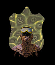
This is one of the army figurines we will be culturing he_la over and infecting with the HIV virus. Testing the possibility of viewing cells directly from the army mens bodies, but no luck...

Soldier silhouettes we will be using as acetate backdrops for animation piece.
Methodology:
The he_la cells were infected with the HIV virus two days prior to the test.
The cells were seeded in one 24 well plate.
Colouration:
Remove medium and add 1 ml of fixing solution (1% formeldehyde, 0.2% glutaraldehyde in PBS) per well for 10 mins at RT (no more than 10 min). Remove fixer and wash twice with PBS. Add staining solution (0.25 mls for 24 well plate, 0.5 ml for 12 well plate) to cells and leave at 37 degrees Celcius. Incubate the plate 2Hr (minimum, we can incubate ON at RT in the dark). The staining reaction is tsopped by washing the plates twice with PBS.
Staining solution should be fresh each time.
For 1 ml
950 microliters PBS
20 microlitres- 0.2 M, Potassium Ferrocyanide
20 microlitres- 0.2 M, Potassium Ferricyanide
10microlitres - 2 M, MgC12
10 microlitres - 40mg/ml X-Gal (make up X-Gal in DMSO and store at -20 degrees Celcius in small aliquots that are protected from the light. The X-Gal stocks will turn yellow over time. This will not affect the assay. Throw away the stocks if the yellow colour turns greenish-brown).
----------------------------------------
To see the virus's growth happen so rapidly was pretty amazing. It really hit home to me just how aggressive this organism is.
It felt really strange to be infecting Henrietta's cells with HIV. Knowing how her family feels about the mass existance of her cells, and what happenined to her makes me feel very uneasy to be using them for this purpose - but, I do feel we are using them for a positive cause, to promote issues relating to those living with HIV, and the phenomenon itself.
In the time that i've spent with her cells and her story, i've come to equate her cell with her body and thus, herself. I guess if you equate tissue with embodied existance, and you consider the massive quantities of her cell line that exist internationally, and all of the various purposes it has been used for, she is in effect a woman who has lived a thousand different lives.

Early morning happiness...


Preparing the stain beforehand in a PC2 Lab.


Entering the AIDS Lab. BLS3 Security.

Checking out he_la cells pre-enzyme.

Washing with PBS Solution.


Staining the he_la cells with the enzyme.

Into the incubator for 2 hours.
After incubating the cells for 1 hour we checked them for visibility and the result was not very encouraging. Although we could see the blue stain very clearly in the infected cells, we could see nothing with the naked eye. Upon checking another hour later though the cells had proliferated rapidly and we could easily see the infected cells in the daylight, without the need for the microscope. Next week when we do this on the he_la cultured army guys we will extend the incubation period and hopefully the cels will proliferate more creating a stronger colour.

After 2 hours incubation the infected cells can be clearly seen in blue.

This is one of the army figurines we will be culturing he_la over and infecting with the HIV virus. Testing the possibility of viewing cells directly from the army mens bodies, but no luck...

Soldier silhouettes we will be using as acetate backdrops for animation piece.
Methodology:
The he_la cells were infected with the HIV virus two days prior to the test.
The cells were seeded in one 24 well plate.
Colouration:
Remove medium and add 1 ml of fixing solution (1% formeldehyde, 0.2% glutaraldehyde in PBS) per well for 10 mins at RT (no more than 10 min). Remove fixer and wash twice with PBS. Add staining solution (0.25 mls for 24 well plate, 0.5 ml for 12 well plate) to cells and leave at 37 degrees Celcius. Incubate the plate 2Hr (minimum, we can incubate ON at RT in the dark). The staining reaction is tsopped by washing the plates twice with PBS.
Staining solution should be fresh each time.
For 1 ml
950 microliters PBS
20 microlitres- 0.2 M, Potassium Ferrocyanide
20 microlitres- 0.2 M, Potassium Ferricyanide
10microlitres - 2 M, MgC12
10 microlitres - 40mg/ml X-Gal (make up X-Gal in DMSO and store at -20 degrees Celcius in small aliquots that are protected from the light. The X-Gal stocks will turn yellow over time. This will not affect the assay. Throw away the stocks if the yellow colour turns greenish-brown).
----------------------------------------
To see the virus's growth happen so rapidly was pretty amazing. It really hit home to me just how aggressive this organism is.
It felt really strange to be infecting Henrietta's cells with HIV. Knowing how her family feels about the mass existance of her cells, and what happenined to her makes me feel very uneasy to be using them for this purpose - but, I do feel we are using them for a positive cause, to promote issues relating to those living with HIV, and the phenomenon itself.
In the time that i've spent with her cells and her story, i've come to equate her cell with her body and thus, herself. I guess if you equate tissue with embodied existance, and you consider the massive quantities of her cell line that exist internationally, and all of the various purposes it has been used for, she is in effect a woman who has lived a thousand different lives.
HIV Project - Staining technique

After much time and energy spent trying to locate a Darkreader transilluminator, or something similar from amny different sources, some of whom had never even heard of one, it was decided that we'd try an alternative technique for cell visualisation, with a blue coloured enzyme which infiltrates infected cells and can be seen with the naked eye. This is a great alternative, as we don't have to worry about locating any new equipment etc as we would with GFP cells, and can take photographic imagery in daylight. Ive never done it before either, so I'm curious to see just how visible the cells will be.
We have our methodology and an appointment for the 29th June, so we are set to go!
Wednesday 27 June 2007
HIV Project Update


Ok today we recieved confirmation from Vrij Uni that we can go ahead with the project, so we are Very Excited!
We will be using he_la cells to culture over glass army figurines, as that is a technique i'm familiar with. There are two options we can go with for the visualisation of the infected cells. The first is to use a blue enzyme which can be seen in daylight with the naked eye. As Lot has not used this technique before she would prefer not to, unless necessary. The second and more favourable option to use is GFP he_la's. To visualise the infected he_la cells effecively however we need to access a transilluminator such as a Darkreader, rather than a straight UV light, which enhances the flourescence substantially, otherwise they will be quite difficult to see, as GFP expression in HIV is quite low. Above is what the Darkreader can do, so it seems to be quite a significant difference.
So right now we are trying to locate a transilluminator which can do this...
Sunday 10 June 2007
A&G Centre Lot de Witte - Meeting #2
Tuesday 5th of June was the the second meeting with Lot de Witte. In the previous meeting with her she implied that it may be possible to actually work with the HIV virus itself - to tissue culture it over 3D glass form, in the Vrij University Immunology Labs. This is really exciting, and in preparation we are organising two small glass army figurines to be ready within a couple of weeks. This meeting with Lot confirmed the possibility of carrying out the project at Vrij, however it has to be cleared with the Dept etc, and it will be Lot who physically works with the virus itself, while we document the process. This is as expected due to the Biological Safety Level required to work with an incurable virus such as HIV/AIDS.
So right now we are waiting to hear back from the Dept, as to whether it gets the go-ahead or not...
So right now we are waiting to hear back from the Dept, as to whether it gets the go-ahead or not...
Subscribe to:
Posts (Atom)